For decades, scientists have understood that the gut hormone GLP-1 plays a crucial role in modern treatments for diabetes and obesity. It is released after a meal when intestinal L cells detect increasing glucose levels. However, the exact mechanism by which glucose signals these cells to release GLP-1 has remained surprisingly unclear.
Now, a team led by Professor Sheng-Cai Lin at Xiamen University has uncovered this long-standing mystery. In a paper published on August 25, 2026, in the journal Vita, the researchers demonstrate that glucose functions not merely as a fuel, but as a direct molecular signal - specifically, a ligand that binds to the enzyme glucokinase (GK) and triggers GLP-1 secretion. This discovery challenges the traditional “metabolic coupling” model, which proposed that glucose must be broken down to produce ATP before GLP-1 can be released.
A Bold Hypothesis Rooted in Basic Biochemistry
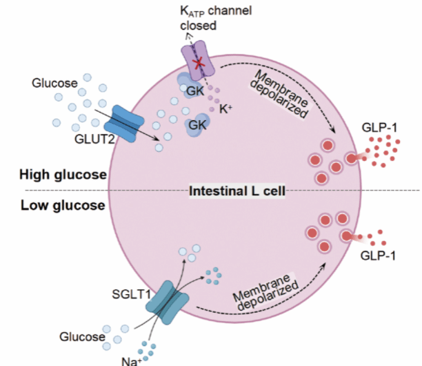
The traditional “metabolic coupling” model is both elegant and intuitive: glucose enters the L cell, is phosphorylated by GK, and then enters glycolysis and the TCA cycle, generating ATP. This increase in ATP leads to the closure of KATP potassium channels, causing the cell membrane to depolarize. As a result, voltage-dependent calcium channels (VDCCs) open, allowing calcium to flow in. This influx of calcium triggers the fusion of GLP-1-containing vesicles with the membrane, resulting in the release of their contents. This model has been used to explain the secretion of insulin, glucagon, and other hormones, and it clarifies why GK activators and KATP blockers stimulate GLP-1 secretion. However, the most fundamental question - whether glucose itself, rather than its metabolic products, actually initiates this process - had not been directly proven.
Professor Lin’s team had a hunch. Their earlier work had revealed a lysosomal glucose-sensing pathway that activates AMPK. In this pathway, low glucose levels are detected not by ATP depletion or decrease, but instead by a reduction in the glycolytic intermediate fructose-1,6-bisphosphate (FBP), which alters the conformation of the enzyme aldolase. This discovery taught them that metabolites can serve as signals in their own right. They then posed the question: Could glucose itself act as a ligand, particularly at the high postprandial concentrations that stimulate GLP-1 secretion?
Biochemistry pointed the way. The four human hexokinases exhibit markedly different affinities for glucose. While hexokinases I through III have Km values below 1 mM, GK has a Km (more precisely, S0.5) greater than 10 mM, aligning with the glucose concentration found in the gut postprandially. Moreover, GLP-1 secretion increases across a wide range of glucose concentrations, with no saturation observed even above 50 mM. These findings led the team to propose that glucose itself is a signaling molecule, with GK acting as its receptor.
Evidence That Glucose Itself Is the Messenger
To investigate this, the researchers used MDGP, a glucose analog that can be taken up and phosphorylated but cannot be metabolized further to produce ATP. They discovered that MDGP still stimulated GLP‑1 release. They also measured ATP levels inside L cells and found no change during high-glucose stimulation, even as GLP-1 secretion increased normally. This indicates that ATP production is not the trigger for GLP-1 secretion in high glucose.
Importantly, blocking both glucose transporters, SGLT1 and GLUT2, abolished GLP-1 secretion, showing that glucose must enter the cell to have an impact on GLP-1 secretion. Interestingly, these two transporters operate in different concentration ranges: SGLT1 functions at low glucose concentrations (below approximately 10 mM) for basal release, while GLUT2 takes over at higher concentrations - specifically, the postprandial range - for the primary secretion of GLP‑1. (SGLT1 also couples sodium transport, providing an alternative, ATP-independent depolarization mechanism, which the team acknowledges as a known parallel pathway.)
GK Acts as a Glucose Receptor
The definitive proof came from studying GK itself. The team found that 2-DG - a glucose analog that can be phosphorylated by GK but does not remain stably bound - failed to stimulate GLP‑1 release. They then examined naturally occurring GK mutants found in patients with maturity-onset diabetes of the young type 2 (MODY2). One mutant, L309P, retains normal glucose binding but has lost catalytic activity. This mutant consistently bound glucose and triggered GLP‑1 release even at low glucose concentrations. Another mutant, G80A, cannot bind glucose and failed to release GLP‑1 even at high glucose levels. These experiments confirmed that it is the binding of glucose to GK, rather than the enzymatic reaction, that drives secretion.
The team also had a unique opportunity to validate this in humans. In collaboration with researchers at Zhongshan Hospital, Fudan University, they studied two MODY2 patients carrying novel GK mutations: one with R397L (which maintains glucose binding) and another with a deletion (Δ92-CT) that prevents binding. Although both patients exhibited similar blood glucose levels after a meal, the patient with the binding-deficient mutation secreted significantly less GLP‑1, providing clinical evidence for the proposed mechanism.
How the Signal Transmits: GK Meets the Potassium Channel
If GK does not alter ATP levels, how does it close the KATP channel? The research team discovered that GK physically binds to the Kir6.2 subunit of the channel, independent of ATP. Through mutagenesis, they identified a critical interaction region on GK, specifically amino acids 205-228. In the glucose-bound state, GK directly inhibits the channel, causing membrane depolarization and GLP-1 release - completely bypassing the traditional ATP-dependent pathway.
A “Double-Lock” Safety Mechanism
This ATP-independent sensing mechanism may hold significant physiological implications. By ensuring that GLP-1 release is strictly dependent on glucose levels, the system prevents false triggers from other nutrients that could raise ATP levels. Additionally, GLP-1 only stimulates insulin secretion from pancreatic β-cells when blood glucose levels are already high. Therefore, even if other nutrients inadvertently increase GLP-1 levels, insulin will not be released inappropriately. This creates a built-in “double-lock” that protects against hypoglycemia.
The team also revisited their earlier work on metformin. They had previously shown that metformin promotes GLP-1 release through the lysosomal AMPK pathway, both at high and low glucose levels. However, the double-lock mechanism ensures that at low glucose levels, the extra GLP-1 does not cause dangerous insulin spikes. This logic is applicable to other lysosomal AMPK pathway agonists as well, such as lithocholic acid.
Clinical Implications for Future Diabetes Therapies
The findings presented in this study open new avenues for drug development. Traditional GK activators (GKAs) encountered issues because they overstimulated the enzyme’s catalytic activity, leading to excessive glycolysis. However, this research shows that GK’s glucose-sensing and catalytic functions can be separated. A potential drug that stabilizes the glucose-bound conformation, without enhancing its catalytic activity, could provide effective treatment with fewer side effects.
Current GLP-1 receptor agonists, such as semaglutide, and DPP-4 inhibitors are highly effective but can cause nausea due to their long-acting nature, which might be excessive for milder cases. A promising direction for precision medicine involves mimicking the body’s native, pulsatile GLP-1 release, thus restoring endogenous secretion rather than solely providing exogenous replacement. This study significantly contributes to our understanding of how this native GLP-1 secretion is triggered. Additionally, the limited capacity of SGLT1 for high-glucose transport highlights the limitations of single SGLT1 inhibitors, suggesting that combination strategies targeting both transporters may prove more effective.
Professor Lin’s research team has successfully completed a long journey. In 2017, they discovered how low glucose levels are sensed via aldolase and FBP, which activates AMPK. Now, they have unveiled how high glucose levels are sensed through GK, which stimulates GLP-1 release. Together, these findings provide a more comprehensive understanding of how cells interpret glucose abundance: not just as fuel, but as a dynamic signal that influences metabolism and physiology.
As Professor Lin said, “This paper is the culmination of my long-standing dream of seeing glucose as a metabolic messenger. Our 2017 work on low-glucose sensing convinced us that metabolites can serve as signals. In this study, when we revisited the fundamental biochemistry of GK’s Km values, everything fell into place. It was not a sudden flash of inspiration; it was the intersection of dreams and accumulated knowledge, the moment when fundamental principles unfolded before our eyes.”
This study involved extensive collaboration with teams from Zhejiang University (Professors Haoxing Xu and Meiqin Hu), Zhongshan Hospital of Fudan University (Professors Xiaoying Li and Lin Zhao), Peking University (Professor Changtao Jiang), Shanghai Sixth People’s Hospital (Professor Teng Ma and Dr. Qingchao Zhu), Xiamen University-affiliated Zhongshan Hospital (Professor Xuehui Hong), and Peking University (Professor Lei Chen). The Analysis and Measurement Center and Laboratory Animal Research Center at Xiamen University also provided essential support. Funding for this research was provided by the National Natural Science Foundation of China and the National Key R&D Program of China.
Link:Glucose acts as a ligand for glucokinase to trigger postprandial secretion of GLP-1
