In a study published in Cell, researchers led by Qiao Wu at Xiamen University identify myosin heavy chain 9 (MYH9) as the cuproptosis executioner and establish that mobilizing endogenous copper stores is sufficient to trigger cuproptosis.
Copper sits at a crossroads of life and death. As an essential cofactor for enzymes driving mitochondrial respiration and metabolism, it is indispensable for cellular function. Yet once intracellular free copper exceeds physiological thresholds, it becomes lethal. The field’s understanding of copper-induced cytotoxicity has evolved from nonspecific oxidative damage—driven by copper-catalyzed reactive oxygen species production—to the concept of cuproptosis. In this paradigm, copper does not act indiscriminately; instead, it targets lipoylated proteins, inducing their aberrant aggregation, destabilizing iron-sulfur cluster proteins, and unleashing a proteotoxic crisis. This conceptual shift recasts copper from a generic chemical toxin to a regulated molecular signal, offering fresh theoretical grounding for copper-overload disorders and new therapeutic perspectives for cancer.
However, two fundamental gaps in the cuproptosis field have persisted. First, does cuproptosis possess an executioner protein, analogous to caspase-3 in apoptosis or gasdermins in pyroptosis? Although several key proteins have been implicated, whether any of them can sense excessive copper and directly execute cuproptosis remains unknown. Second, can endogenous copper at physiological concentrations initiate cuproptosis? Prior studies have relied on copper ionophores or supraphysiological doses of exogenous copper to force intracellular overload, but the pathophysiological relevance of such artificial copper stress is questionable. Systemic copper accumulation also risks severe hepatotoxicity and nephrotoxicity, creating a formidable barrier to clinical translation. Thus, whether mobilizing intracellular copper stores alone—without increasing systemic copper burden—can initiate cuproptosis remains a critical question.
On September 14, 2026, the group led by Professor Qiao Wu at the School of Life Sciences, Xiamen University, published a study in Cell entitled “Copper ion-induced MYH9 polymerization executes cuproptosis.” The team identified, for the first time, myosin heavy chain 9 (MYH9) as the executioner of cuproptosis. Using MYH9 as a target, they screened and identified HThPA, the first non-copper-ionophore small-molecule compound capable of inducing copper release from intracellular stores, thereby effectively initiating the cuproptosis program. The study further revealed that drug-resistant tumors, owing to their elevated copper burden, exhibit heightened sensitivity to endogenous copper-mediated cuproptosis, opening a new avenue for overcoming drug-resistant tumors through intracellular endogenous copper-dependent cuproptosis.
Polymerization of lipoylated proteins such as DLAT is intimately linked to cuproptosis. To systematically identify proteins that sense copper and assemble into polymers, the team pursued two parallel strategies: treating live cells with copper ionophores (DSF or ES) and incubating whole-cell lysates directly with copper in vitro. After enriching copper-binding proteins and resolving high-molecular-weight complexes by non-reducing SDS-PAGE, mass spectrometry revealed MYH9 as a standout candidate.
How does MYH9 transduce the copper signal? The study showed that copper ions directly bind to and induce MYH9 polymerization in a dose-dependent manner. Copper ionophore treatment or in vitro copper incubation robustly induced MYH9 polymerization, whereas copper chelators completely blocked this process. To establish causality, the authors assembled three lines of evidence: first, MYH9 knockout suppressed cuproptosis, establishing MYH9 as a key node in the pathway. Second, MYH9 polymerization preceded cell death at lower doses, indicating that it is an upstream trigger rather than a downstream consequence. Most compellingly, optogenetic induction of MYH9 polymerization—in the complete absence of copper—was sufficient to trigger cuproptosis. The generality of this mechanism extends beyond pharmacological models: in acute myeloid leukemia cells, heme deficiency-induced cuproptosis also required MYH9 polymerization, underscoring its physiological relevance.
How does MYH9 drive cuproptosis mechanistically? Distinct forms of cell death often exhibit unique morphological signatures. Across more than a dozen tumor cell lines exposed to copper overload, cells consistently lost their spread morphology and retracted into spherical shapes—a hallmark of cytoskeletal collapse. Indeed, copper binding to MYH9 facilitates the recruitment of actin to MYH9, thereby blocking the assembly of globular actin (G-actin) into F-actin, leading to cytoskeletal collapse and execution of cuproptosis.
Notably, MYH9 comprises 1,960 amino acid residues with a high abundance of basic amino acids (207 lysine, 123 arginine, and 28 histidine residues) and has a positively charged surface that creates electrostatic repulsion against positively charged copper ions—an evolutionary failsafe against accidental activation. So how do cells overcome this barrier at relatively low intracellular copper concentrations? The answer lies in a molecular key. Copper ionophore treatments rupture mitochondrial membranes, releasing DLAT into the cytosol. Cytosolic DLAT binds MYH9 and deploys a non-canonical acetyltransferase activity, acetylating lysine residues to neutralize their positive charge. This local reduction in surface electrostatic potential effectively unlocks MYH9, permitting copper binding. This process represents an elegant “dual-lock” design: electrostatic repulsion prevents inadvertent activation, while DLAT-mediated acetylation provides a sensitization mechanism that unlocks the system when death signals arrive.
Copper ionophores can induce cuproptosis by importing exogenous copper into tumor cells, but their efficacy is highly dose-dependent and systemic copper accumulation causes severe organ toxicity, thereby limiting the translational prospects of cuproptosis-based therapy. To bypass systemic toxicity, Wu’s team reasoned that tumor cells, which often harbor elevated copper levels, might carry sufficient endogenous stores to fuel their own destruction. They therefore established an ATP-based screening platform to identify MYH9-dependent cell death inducers that require no extracellular copper. From an in-house library of 1,163 small molecules, they identified HThPA—the first non-copper-ionophore cuproptosis inducer. Unlike copper ionophores, HThPA does not increase total intracellular copper levels by transporting exogenous copper; instead, it activates endogenous free copper to induce MYH9 polymerization and cuproptosis. Mechanistically, HThPA binds to and stabilizes the conformation of the nuclear receptor Nur77, generating an interface that recruits PDK1. Acting as a scaffold, Nur77 positions PDK1 to phosphorylate and activate CHAC1, which rapidly degrades glutathione (GSH). Because GSH sequesters a major fraction of intracellular copper, its degradation liberates free copper to bind MYH9, whose polymerization further drives cuproptosis.
Finally, the research team elucidated the pathological significance and translational potential of endogenous copper-mediated cuproptosis. In drug-resistant tumor cell lines and in clinical specimens of hepatocellular and gastric carcinoma, resistant tumors consistently displayed higher copper burdens than their drug-sensitive counterparts. This metabolic vulnerability rendered them exquisitely sensitive to HThPA. In drug-resistant xenografts, patient-derived xenograft (PDX) models, and primary liver and gastric cancer models, HThPA potently suppressed tumor growth—particularly in resistant settings—with toxicity profiles nearly indistinguishable from those of vehicle controls, reflecting a substantially wider therapeutic window than ES-Cu-based regimens. This discovery moves beyond the field’s long-standing reliance on copper ionophores, showing that cuproptosis can be initiated not only by exogenous copper overload but also by actively perturbing endogenous copper homeostasis. Endogenous copper release, the study argues, is not a passive metabolic byproduct but an actionable signal that determines cell fate.
In summary, these findings answer the two defining questions of the cuproptosis field. They identify MYH9 as the executioner of cuproptosis, elucidate the mechanism by which copper binding induces MYH9 polymerization and consequent disruption of the actin cytoskeleton, and propose MYH9 polymerization as a potential biomarker for cuproptosis. They further reveal a complete endogenous copper-mediated cuproptosis pathway and deliver a first-in-class tool compound that reverses drug resistance without systemic copper toxicity.
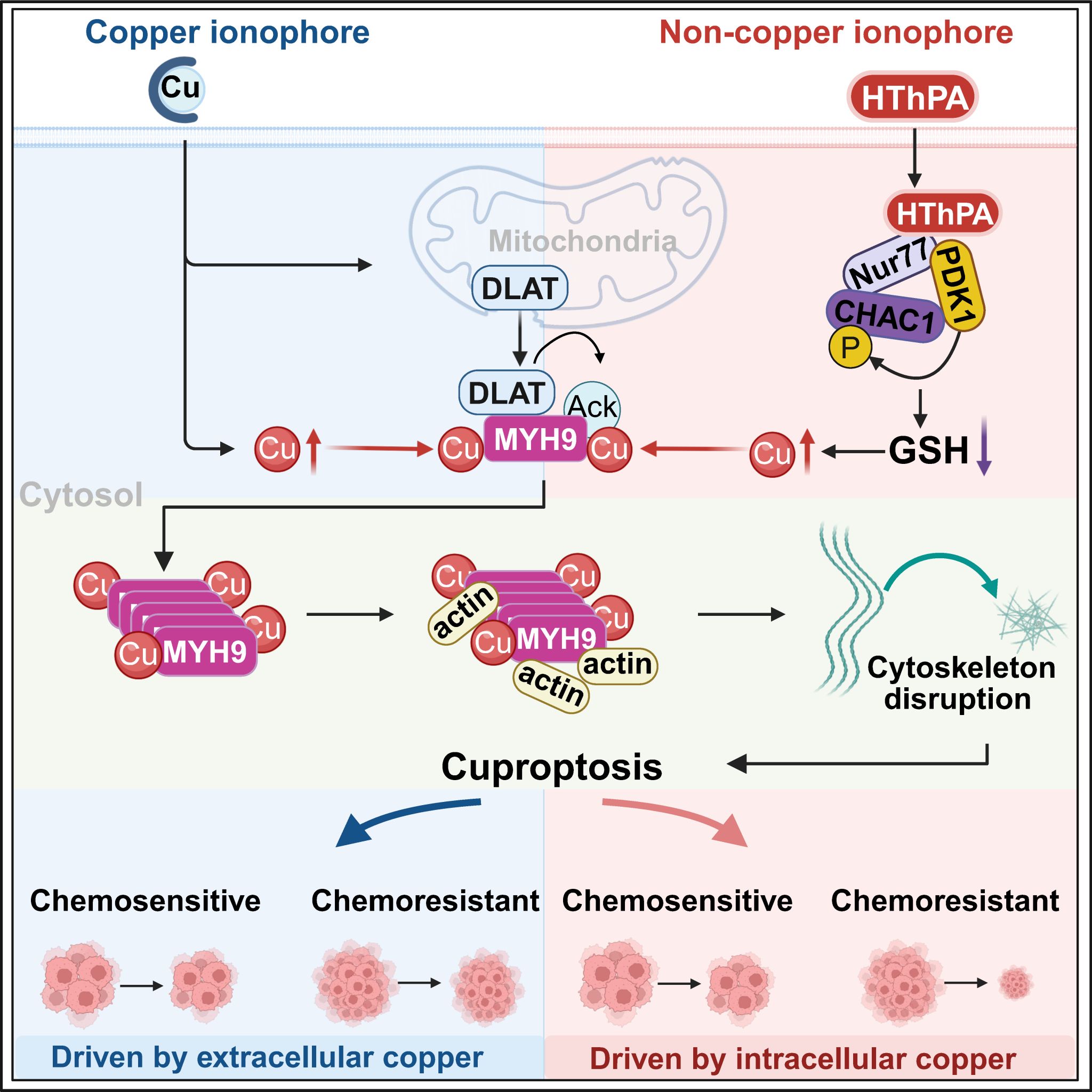
Working model: From “Exogenous Overload” to “Endogenous Unlocking” MYH9 as the executioner protein integrates exogenous and endogenous copper signals. Exogenous copper enters cells via ionophores, while HThPA triggers the Nur77-PDK1-CHAC1 axis to degrade GSH and mobilize endogenous copper stores. In both scenarios, DLAT-mediated acetylation sensitizes MYH9 by neutralizing its electrostatic shield, enabling copper binding, polymerization, and cytoskeletal collapse that executes cuproptosis.
For decades, copper was cast in a binary role: metabolic essential or indiscriminate poison. This work reveals a far more sophisticated reality. At the cellular crossroads of survival and death, copper becomes neither nutrient nor toxin, but a precisely regulated signal—one that can be read, redirected, and weaponized against cancer. In reframing copper from a toxin to be expelled to a signal to be harnessed, the study opens new avenues for treating copper-overload disorders such as Wilson’s disease and, critically, offers a mechanistic blueprint for overcoming one of oncology’s most stubborn challenges: therapeutic resistance.
Professors Qiao Wu, Hangzi Chen, Xuehui Hong, Tianwei Lin, and Associate Professor Funan Li of Xiamen University are co-corresponding authors of this paper. Doctoral students Liuzheng Wu, Xiangyu Mi, and Zaijun Liu (School of Life Sciences), Yang Ding (School of Pharmaceutical Sciences), and postdoctoral fellow Wenbin Hong (First Affiliated Hospital of Xiamen University) are co-first authors. This project was supported by multiple national funding agencies.
Link: https://doi.org/10.1016/j.cell.2026.08.030
